
Radiology
Radiology is the medical discipline that uses medical imaging to diagnose and treat diseases within the bodies of animals, including humans.
Radiologists are medical doctors that specialize in diagnosing and treating injuries and diseases using medical imaging (radiology) procedures ( exams/tests) such as X-rays, computed tomography (CT), magnetic resonance imaging (MRI), nuclear medicine, positron emission tomography (PET) and ultrasound.
Advanced Detection and Analysis with Radiology
Radiology plays several crucial roles in medicine. It can be used for advanced testing and treatment, for screening and wellness, and to detect diseases and conditions. Think of radiology as a huge umbrella, with many specific technologies underneath it.
Cone-beam computed tomography systems (CBCT),is a radiographic imaging method that allows accurate, three-dimensional (3D) imaging of hard tissue structures. CBCT is the most significant among the medical diagnostic imaging modalities that have emerged recently.
Modle: Pax-i3D provides 4 multi FOV sizes ranging from 5x5 to 12x9. By selecting the appropriate FOV size, you can have the optimum image for your diagnostic needs reducing unnecessary X-ray radiation for patients.

X-ray, or Radiography, is used to diagnose fractured bones, detect injury or infection, or to locate foreign objects in soft tissue. Some x-ray exams employ an iodine-based contrast material to clarify the visibility of specific organs like the heart, lungs, blood vessels or tissues.
They're mainly used to look at the bones and joints, although they're sometimes used to detect problems affecting soft tissue, such as internal organs. Problems that may be detected during an X-ray include: bone fractures and breaks.

Computed Tomography (CT) creates detailed images of internal organs, bones, soft tissue and blood vessels. CT is considered by many doctors as the preferred method to detect cancer, since it can confirm the presence of a tumor and determine its size and location. In emergency cases, CT can quickly reveal internal injuries and bleeding to help save lives.
Magnetic Resonance Imaging (MRI)uses a powerful magnetic field and radio waves to detect conditions such as tumors and diseases of the liver, heart and bowel. MRI may also be used to monitor an unborn child in the womb.
Modle: Hitachi aperto 0.4T MRI provides 4 multi FOV sizes ranging from 5x5 to 12x9. By selecting the appropriate FOV size, you can have the optimum image for your diagnostic needs reducing unnecessary X-ray radiation for patients.

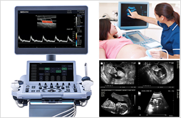
Ultrasound Imagingis an effective method of diagnosing unexplained pain, swelling and infection. It can provide imaging guidance for needle biopsies or evaluate conditions related to blood flow. Ultrasound is also the preferred imaging method for monitoring a pregnant woman and her unborn child.
Modle: Edan Acclarix LX9 developed specifically to address the challenges of complex ultrasound environments, the Acclarix LX9 Diagnostic Ultrasound System aims to provide precise diagnosis in versatile clinic application with uncompromising image quality , brilliant workflow and user-friendly operation.
Mammography Breast Imaging uses low-dose x-rays to detect cancer early —when it is most treatable. Mammography plays a leading role in early detection, because it can show changes in the breast up to two years before you or your physician can feel them.